Within Harris Birthright there is now a unit dedicated
specifically to detailed examination of the fetal heart. The fetal cardiac unit integrates
skills and experience in congenital heart disease with those in fetal medicine to optimise
the detection and management of heart abnormalities in the fetus, whether they be primary
abnormalities of the heart or disturbances of heart function related to other fetal
conditions.
- What is congenital heart disease?
Malformations of the heart account for the majority of
heart disease in the fetus and in children. There are a wide variety of individual
conditions ranging from simple holes connecting heart chambers to complex abnormalities in
which one of the heart chambers is effectively absent or vessels and chambers are
abnormally connected or obstructed. Less common are abnormalities in the heart rhythm,
where the heart rate is too fast, too slow, or irregular and abnormalities of the heart
muscle, in which the heart fails to contract normally.
- Why scan the fetal heart?
Structural congenital heart disease is relatively common,
occurring in about 8 per thousand live-born babies. Of these, about 5 per thousand could
be identified in the fetus by expert level fetal heart scanning. Moreover, those heart
abnormalities that can be detected pre-natally are the more severe ones, with the most
profound influence on the survival and well being of the child.
Abnormalities of the heart often occur as part of
conditions such as Down’s syndrome and many other conditions associated with mental
retardation. An examination of the fetal heart is therefore an essential part of the
complete evaluation when any abnormality has been recognised on a scan. Conversely, the
reassurance derived from a normal detailed anomaly scan is only fully substantiated if the
fetal heart has been examined in detail.
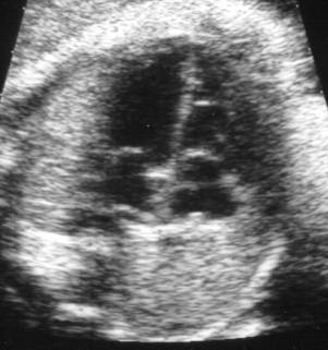
Scan of a fetal heart
- Why is a special unit
necessary?
Most hospitals offer routine ultrasound screening at about
20 weeks, which usually includes examination of a simple 4-chamber view of the heart and
may identify some, but not all, major heart abnormalities. In many serious heart defects
however, the 4-chamber view of the heart is entirely normal. Therefore a more detailed
examination of the heart, including the connections of the major blood vessels and Doppler
estimation of flow through the valves, is necessary to be confident about excluding most
significant abnormalities. It is therefore appropriate that those at increased risk of
having a fetus with a congenital heart abnormality should be offered a specialist fetal
heart scan. Also, when an abnormality of the heart is suspected on a routine anomaly scan
the abnormality should be confirmed and precisely defined in a specialist centre.
Advances in cardiac surgery over recent years mean that
there are now only a few rare congenital heart abnormalities for which no treatment can be
offered. The outlook for various types of abnormality is however very different. Small but
important details in complex abnormalities can have a major influence on the type of
surgery possible and the likelihood of success. In addition to detailed analysis of the
structure and function of an abnormal fetal heart it is therefore important to use this
information to provide prospective parents with an accurate prediction of the likely
outlook for their babies condition. In this respect, it is not only the chance of survival
that is important, but also the quality of life that may be expected for survivors. Close
and continuing links with the paediatric cardiology department at Guy’s, including
weekly meetings with the cardiac surgeons to discuss cases, ensures that the fetal
cardiology unit at King’s remains in tune with new developments in treatment.
- Who requires a specialist
fetal heart scan?
Certain women can be identified as being at higher than
average risk of having a fetus affected by congenital heart disease for the following
reasons.
- Suspicion of a heart anomaly on a routine scan, or failure
to adequately demonstrate a normal 4-chamber view of the heart on a routine anomaly scan.
- Increased thickness of nuchal fluid on an early (12-week)
scan.
- Either parent or another close relative having congenital
heart disease.
- Diabetes in the mother.
- Exposure of the fetus to drugs such as lithium,
anticonvulsants or alcohol in early pregnancy.
- Demonstration of any extra-cardiac abnormality in the fetus,
such as exomphalos or cleft palate, that might conceivably be part of a syndrome.
- Abnormal fetal heart rhythm.
- Multiple pregnancy
However, most congenital heart disease occurs in women in
whom there are no reasons to expect an increased risk. It has therefore been our policy at
Harris Birthright to ensure that a detailed fetal heart scan is performed by a doctor with
special experience and expertise in every woman who attends for detailed fetal anomaly
scanning, even in the absence of any identifiable risk factors.
|